
 University of Edinburgh
Benjamin Jin is a doctoral researcher within the Medical Research Council's Precision Medicine Doctoral Training Programme at the University of Edinburgh. He holds bachelor's and master's degrees in computer science from the University of Augsburg, Germany. His research focuses on medical image analysis of routine clinical imaging for the prevention of neurovascular disease.
Education
-
University of EdinburghInstitute for Neuroscience and Cardiovascular Research
Doctoral ResearcherSep. 2023 - present -
 University of AugsburgM.Sc. in Computer ScienceOct. 2019 - Aug. 2022
-
University of AugsburgB.Sc. in Computer ScienceOct. 2016 - Oct. 2019
Experience
-
 Erasmus MCVisiting ResearcherJan. 2026 - Feb. 2026
-
 Recoro UGSoftware Engineer and Data ScientistJan. 2022 - Feb. 2023
-
University of AugsburgResearch and Teaching AssistantNov. 2020 - Sep. 2021
-
 Meteocontrol GmbHSoftware DeveloperJan. 2022 - Feb. 2023
-
 itestra GmbHSoftware DeveloperAug. 2017 - Oct. 2017
Honors & Awards
-
SINAPSE ECR Exchange Fund (UK)2025
-
MRC doctoral studentship (UK)2023-2027
-
EXIST Business Startup Grant (DE)2022
-
Deutschlandstipendium (DE)2017-2021
News
Selected Publications (view all )
Automated Distinction of Intimal and Medial Intracranial Arterial Calcification from CT Head
Benjamin Jin, Maria del C. Valdés Hernández, Richard Bortsov, Joanna M. Wardlaw, Daniel Bos, Grant Mair
Accepted at SWITCH+ @ MICCAI 2026arXiv 2026
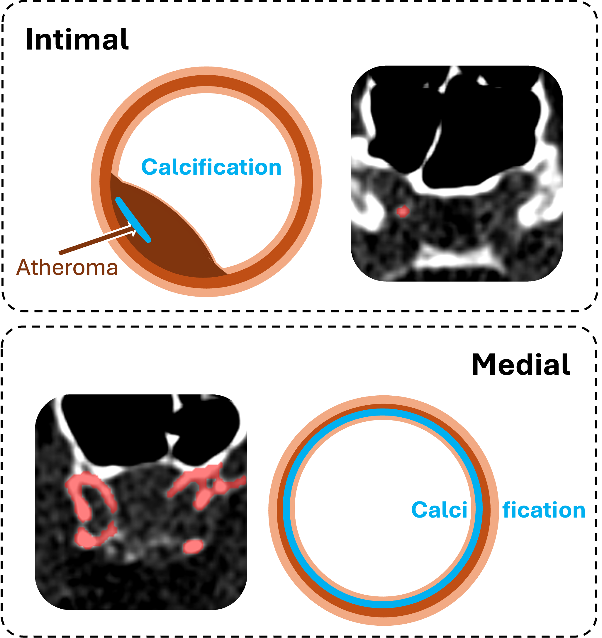
Intracranial arterial calcifications (IACs) are a common finding on clinical non-contrast enhanced head CT scans and are associated with neurovascular disease. Calcifications can occur in the intimal or medial layer of the arterial wall, subtypes that differ in aetiology and may have distinct clinical relevance. These subtypes can be visually distinguished by radiologists based on the shape of the calcifications. We investigate three automated approaches for subtype classification of IAC from head CT-derived segmentation masks: (1) an automated adaptation of the established radiological visual score, (2) a sphericity-based method, and (3) a method based on shape embeddings extracted by a medical shape foundation model. All approaches use the same lightweight classification pipeline on top of the features they compute and are evaluated using 5-fold cross-validation. The three methods achieved comparable performance, with the embedding-based approach yielding the best overall results with a weighted F1 (mean ± SD) of up to 71.5 ± 3.7 for a single artery and 59.8 ± 1.7 for the joint artery classification. Performance was largely preserved when using automated instead of manual IAC segmentation masks, and we found the difference in weighted F1 not significant. Our results show that fully automated IAC subtype quantification from head CT is feasible and remains robust to the use of manual and automated IAC segmentation masks.
Automated Distinction of Intimal and Medial Intracranial Arterial Calcification from CT Head
Benjamin Jin, Maria del C. Valdés Hernández, Richard Bortsov, Joanna M. Wardlaw, Daniel Bos, Grant Mair
Accepted at SWITCH+ @ MICCAI 2026arXiv 2026
Intracranial arterial calcifications (IACs) are a common finding on clinical non-contrast enhanced head CT scans and are associated with neurovascular disease. Calcifications can occur in the intimal or medial layer of the arterial wall, subtypes that differ in aetiology and may have distinct clinical relevance. These subtypes can be visually distinguished by radiologists based on the shape of the calcifications. We investigate three automated approaches for subtype classification of IAC from head CT-derived segmentation masks: (1) an automated adaptation of the established radiological visual score, (2) a sphericity-based method, and (3) a method based on shape embeddings extracted by a medical shape foundation model. All approaches use the same lightweight classification pipeline on top of the features they compute and are evaluated using 5-fold cross-validation. The three methods achieved comparable performance, with the embedding-based approach yielding the best overall results with a weighted F1 (mean ± SD) of up to 71.5 ± 3.7 for a single artery and 59.8 ± 1.7 for the joint artery classification. Performance was largely preserved when using automated instead of manual IAC segmentation masks, and we found the difference in weighted F1 not significant. Our results show that fully automated IAC subtype quantification from head CT is feasible and remains robust to the use of manual and automated IAC segmentation masks.

Calibrated Self-supervised Vision Transformers Improve Intracranial Arterial Calcification Segmentation from Clinical CT Head Scans
Benjamin Jin, Grant Mair, Joanna M. Wardlaw, Maria del C. Valdés Hernández
Data Engineering in Medical Imaging @ Medical Image Computing and Computer Assisted Interventions (MICCAI) 2025
Vision Transformers (ViTs) have gained significant popularity in the natural image domain but have been less successful in 3D medical image segmentation. Nevertheless, 3D ViTs are particularly interesting for large medical imaging volumes due to their efficient self-supervised training within the masked autoencoder (MAE) framework, which enables the use of imaging data without the need for expensive manual annotations. intracranial arterial calcification (IAC) is an imaging biomarker visible on routinely acquired CT scans linked to neurovascular diseases such as stroke and dementia, and automated IAC quantification could enable their large-scale risk assessment. We pre-train ViTs with MAE and fine-tune them for IAC segmentation for the first time. To develop our models, we use highly heterogeneous data from a large clinical trial, the third International Stroke Trial (IST-3). We evaluate key aspects of MAE pre-trained ViTs in IAC segmentation, and analyse the clinical implications. We show: 1) our calibrated self-supervised ViT beats a strong supervised nnU-Net baseline by 3.2 Dice points, 2) low patch sizes are crucial for ViTs for IAC segmentation and interpolation upsampling with regular convolutions is preferable to transposed convolutions for ViT-based models, and 3) our ViTs increase robustness to higher slice thicknesses and improve risk group classification in a clinical scenario by 46%.
Calibrated Self-supervised Vision Transformers Improve Intracranial Arterial Calcification Segmentation from Clinical CT Head Scans
Benjamin Jin, Grant Mair, Joanna M. Wardlaw, Maria del C. Valdés Hernández
Data Engineering in Medical Imaging @ Medical Image Computing and Computer Assisted Interventions (MICCAI) 2025
Vision Transformers (ViTs) have gained significant popularity in the natural image domain but have been less successful in 3D medical image segmentation. Nevertheless, 3D ViTs are particularly interesting for large medical imaging volumes due to their efficient self-supervised training within the masked autoencoder (MAE) framework, which enables the use of imaging data without the need for expensive manual annotations. intracranial arterial calcification (IAC) is an imaging biomarker visible on routinely acquired CT scans linked to neurovascular diseases such as stroke and dementia, and automated IAC quantification could enable their large-scale risk assessment. We pre-train ViTs with MAE and fine-tune them for IAC segmentation for the first time. To develop our models, we use highly heterogeneous data from a large clinical trial, the third International Stroke Trial (IST-3). We evaluate key aspects of MAE pre-trained ViTs in IAC segmentation, and analyse the clinical implications. We show: 1) our calibrated self-supervised ViT beats a strong supervised nnU-Net baseline by 3.2 Dice points, 2) low patch sizes are crucial for ViTs for IAC segmentation and interpolation upsampling with regular convolutions is preferable to transposed convolutions for ViT-based models, and 3) our ViTs increase robustness to higher slice thicknesses and improve risk group classification in a clinical scenario by 46%.

Pre-processing and Quality Control of Large Clinical CT Head Datasets for Intracranial Arterial Calcification Segmentation
Benjamin Jin, Maria del C. Valdés Hernández, Alessandro Fontanellea, Wenwen Li, Eleanor Platt, Paul Armitage, Amos Storkey, Joanna M. Wardlaw, Grant Mair
Data Engineering in Medical Imaging @ Medical Image Computing and Computer Assisted Interventions (MICCAI) 2024
As a potential non-invasive biomarker for ischaemic stroke, intracranial arterial calcification (IAC) could be used for stroke risk assessment on CT head scans routinely acquired for other reasons (e.g. trauma, confusion). Artificial intelligence methods can support IAC scoring, but they have not yet been developed for clinical imaging. Large heterogeneous clinical CT datasets are necessary for the training of such methods, but they exhibit expected and unexpected data anomalies. Using CTs from a large clinical trial, the third International Stroke Trial (IST-3), we propose a pipeline that uses as input non-enhanced CT scans to output regions of interest capturing selected large intracranial arteries for IAC scoring. Our method uses co-registration with templates. We focus on quality control, using information presence along the z-axis of the imaging to group and apply similarity measures (structural similarity index measure) to triage assessment of individual image series. Additionally, we propose superimposing thresholded binary masks of the series to inspect large quantities of data in parallel. We identify and exclude unrecoverable samples and registration failures. In total, our pipeline processes 10,659 CT series, rejecting 4,322 (41%) in the entire process, 1,450 (14% of the total) during quality control, and outputting 6,337 series. Our pipeline enables effective and efficient region of interest localisation for targeted IAC segmentation.
Pre-processing and Quality Control of Large Clinical CT Head Datasets for Intracranial Arterial Calcification Segmentation
Benjamin Jin, Maria del C. Valdés Hernández, Alessandro Fontanellea, Wenwen Li, Eleanor Platt, Paul Armitage, Amos Storkey, Joanna M. Wardlaw, Grant Mair
Data Engineering in Medical Imaging @ Medical Image Computing and Computer Assisted Interventions (MICCAI) 2024
As a potential non-invasive biomarker for ischaemic stroke, intracranial arterial calcification (IAC) could be used for stroke risk assessment on CT head scans routinely acquired for other reasons (e.g. trauma, confusion). Artificial intelligence methods can support IAC scoring, but they have not yet been developed for clinical imaging. Large heterogeneous clinical CT datasets are necessary for the training of such methods, but they exhibit expected and unexpected data anomalies. Using CTs from a large clinical trial, the third International Stroke Trial (IST-3), we propose a pipeline that uses as input non-enhanced CT scans to output regions of interest capturing selected large intracranial arteries for IAC scoring. Our method uses co-registration with templates. We focus on quality control, using information presence along the z-axis of the imaging to group and apply similarity measures (structural similarity index measure) to triage assessment of individual image series. Additionally, we propose superimposing thresholded binary masks of the series to inspect large quantities of data in parallel. We identify and exclude unrecoverable samples and registration failures. In total, our pipeline processes 10,659 CT series, rejecting 4,322 (41%) in the entire process, 1,450 (14% of the total) during quality control, and outputting 6,337 series. Our pipeline enables effective and efficient region of interest localisation for targeted IAC segmentation.